MicroCT Facility

The USU MicroCT Facility is associated with the Department of Biology and the USU Office of Research. The facility opened in September of 2022, and houses a Nikon XT H 225 ST 2x µCT scanner and two state of the art workstations with VG Studio Max installed. The acquisition of this equipment was funded in part by a National Science Foundation Major Instrumentation grant, along with additional funds contributed by the Office of Research, the College of Arts & Sciences, the College of Agriculture and Applied Sciences, the Agricultural Experiment Station, and the departments of Biology, ADVS, and Geosciences.

Checklist Icon

Scheduling & Calendar

Checklist Icon

Using the Facility

Facility Information
Location: ESLC 055A
Email: microCT@usu.edu 

Availability by Appointment: 9 a.m.- 5 p.m. M-F
*Trained users may use the facility after hours and on weekends

Facility Staff

Why MicroCT?

MicroCT scanning allows users to collect a series of 2D projections as the sample is rotated and to reconstruct these projections to create a 3D volume. This volume can then be digitally manipulated to resolve micron-level details of a given sample, allowing users to examine both the external and internal structure/composition non-destructively. Thus, microCT can be a valuable tool for a wide range of disciplines.

What Can You Scan?

X-ray icon Biological/Organic

  • Mineralized material (e.g., bone, shells, etc.) can be scanned without any additional preparation. 
  • Non-mineralized material (e.g., plant roots, chitinous exoskeletons, and soft tissues) must be stained with a contrast agent prior to staining. 

Rockslide icon Geological

  • Rocks
  • Minerals
  • Core samples

Gear icon Engineering

  • Metalic parts
  • Machine parts

Capabilities & Specifications
  • Tungsten target, two interchangeable target heads
    • 225kV Reflection target with focal spot size down to 3 microns
    • 180kV Transmission target with focal spot size down to 1 micron
  • Maximum 225kV for high absorbing samples (e.g., metallic samples, dense minerals)
  • Maximum frame rate of 30fps
  • Maximum sample size:
    • Maximum dimensions: At the lowest magnification, the dimensional limits for a given sample are 450mm width by 350mm height (roughly the size of a domestic cat). However, it is possible to perform piecewise scans of larger samples and stitch them together following reconstruction of the volumes
    • Maximum weight: 50kg (110lbs.)
  • Scan types:
    • Circular scan (full 360 rotation)
    • Half-turn scan
    • Tall scan (multi-part scans of large samples that exceed the vertical FOV)
    • Batch scan
    • 2D radiography

News

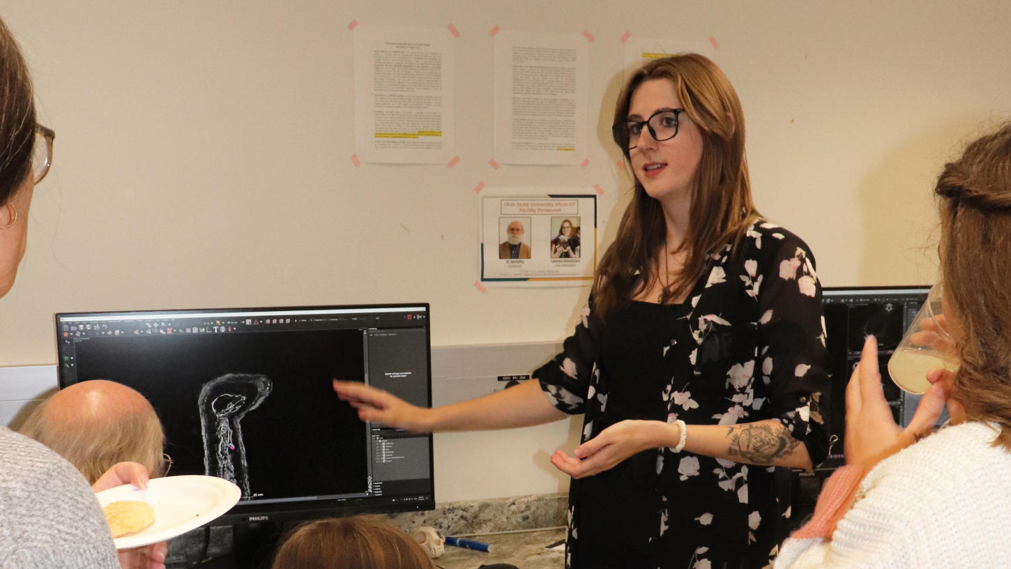
USU's Science Unwrapped Explores 'Mummy Mystery'

What do you do when you (literally) can’t unwrap a scientific mystery? Utah State University anthropologists Sascha Baldauf and Molly Cannon faced this puzzle, when the USU Museum of Anthropology came into possession of ancient mummified remains.

Logan Hammer Studies Varied Snake Species

Undergrad researcher Logan Hammer, left, studies fang length, gape size in varied snake species with mentors, from left, Professor Al Savitzky and graduate student Lauren Houstoun Craige, Utah State University.

Publications